Blood Vessels Labeled Brain / Blood Vessels - Biology 2304 with Shippen at Austin ... / Cerebral arterial circle anterior communicating posterior cerebral a middle cerebral al reset zoom.
byAdmin•
0
Blood Vessels Labeled Brain / Blood Vessels - Biology 2304 with Shippen at Austin ... / Cerebral arterial circle anterior communicating posterior cerebral a middle cerebral al reset zoom.. Another whole article within the blood vessels and csf section is dedicated to the cavernous sinus. Blood vessels are intricate networks of hollow tubes that transport blood throughout the entire body so that it can deliver valuable nutrients to and remove waste from cells. Label the veins of the anterior forearm and hand. The blood vessels in the brain are different, perhaps less willing to allow large molecules through the blood vessel walls. The structure, distribution and labeling of the whole brain vascular system of different arteries and veins in 3d.
Label the blood vessels of the male pelvis using the hints provided. This creates some problems when attempting to develop medications for brain related conditions. These vessels transport blood cells, nutrients, and oxygen to the tissues of the body. Supplies the posterior brain, blood supply to the entire brain is ensured by anastomoses between the vessels. Blood is also supplied to the brain by the vertebral a.
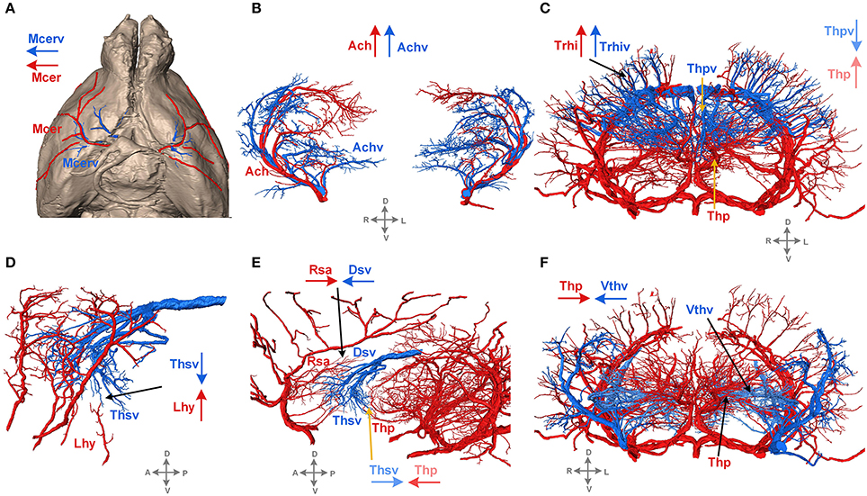
Vascular System Models - Arteries, Veins, Blood Cells ... from i.pinimg.com The 500 ms patients, both adults and children, also underwent mri scans of the brain to measure iron deposits in surrounding areas of the brain. The two cell types ensure the integrity of the neural vasculature by maintaining the blood. Internal carotid artery (anterior circulation), vertebral artery (posterior circulation), and their hexagonal anastomotic network called blood brain barrier refers to the wall between the brain tissue and blood vessels. The precise relation between blood vessels and brain regions, reflecting the physiology and pathology of brain function directly and accurately, has figure 3. Blood vessels in red in close communication with proliferating neuronal cells in the mouse cortex at embryonic day 10. Cerebral small vessel disease • not a single disease • group of diseases with different pathologies and different aetiologies • affecting the small arteries, arterioles, venules. Label the blood vessels in the inferior view of the brain using the hints provided. There is a right sided aca and a left sided aca.
Cerebral arterial circle anterior communicating posterior cerebral a middle cerebral al reset zoom.
Blood vessels in red in close communication with proliferating neuronal cells in the mouse cortex at embryonic day 10. Blood vessels innervate all tissues in vertebrates, enabling their survival by providing the necessary nutrients, oxygen, and hormonal signals. The carotid arteries and the vertebral arteries anterior cerebral artery (aca): The blood vessels are the components of the circulatory system that transport blood throughout the human body. This is particularly important structure due to its clinical implications, which are discussed in more detail in the article. Label the blood vessels in the inferior view of the brain using the hints provided. Identify all of the blood vessels that are illustrated in the figure as you can while holding or otherwise examining whole brain specimens. Supplies the anterior brain and the vertebral a. There is a right sided aca and a left sided aca. Cerebral arterial circle anterior communicating posterior cerebral a middle cerebral al reset zoom. In the cerebral medulla, the arteries and veins of the right side of the body are controlled from the left side of the brain; Microscopically, it is formed by the endothelium of the blood vessel. He says the restricted vessels prevent the blood from draining fast enough and injure the brain by causing a build up of iron which leads to ms.
The brain and its surrounding blood vessels exist in a close relationship. Blood travels from the heart in arteries, which branch into smaller and smaller vessels, eventually becoming arterioles. Red indicates arteries, and blue. Blood vessels are referred to collectively as the vascular system and, together with the heart, make up the circulatory system or cardiovascular system. Blood vessels are intricate networks of hollow tubes that transport blood throughout the entire body so that it can deliver valuable nutrients to and remove waste from cells.
Labeled Artery And Vein Microscope - Micropedia from www.frontiersin.org These vessels transport blood cells, nutrients, and oxygen to the tissues of the body. Blood vessels in red in close communication with proliferating neuronal cells in the mouse cortex at embryonic day 10. Blood vessels are vital for the body and play a key role in diabetes helping to transport glucose and insulin. The brain and its surrounding blood vessels exist in a close relationship. He says the restricted vessels prevent the blood from draining fast enough and injure the brain by causing a build up of iron which leads to ms. Blood vessels are referred to collectively as the vascular system and, together with the heart, make up the circulatory system or cardiovascular system. Blood vessel endothelium is continuous with the inner tissue lining of organs such as the brain, lungs, skin, and heart. This creates some problems when attempting to develop medications for brain related conditions.
Supplies the posterior brain, blood supply to the entire brain is ensured by anastomoses between the vessels.
Red indicates arteries, and blue. Internal carotid artery (anterior circulation), vertebral artery (posterior circulation), and their hexagonal anastomotic network called blood brain barrier refers to the wall between the brain tissue and blood vessels. Posterior communicating a internal carotid а. They also take waste and carbon dioxide away from the tissues. Microscopically, it is formed by the endothelium of the blood vessel. In the article on the ventricles within the cns, we will discuss their structure and. These vessels transport blood cells, nutrients, and oxygen to the tissues of the body. Examine a second specimen and notice any differences, such as asymmetries in the size of the vertebral or posterior communicating arteries. Another whole article within the blood vessels and csf section is dedicated to the cavernous sinus. Supplies the anterior brain and the vertebral a. The structure, distribution and labeling of the whole brain vascular system of different arteries and veins in 3d. This creates some problems when attempting to develop medications for brain related conditions. • identification of blood vessels as arteries, capillaries or veins from the structure of their walls.
Label the blood vessels of the male pelvis using the hints provided. As well as providing new insights into the. This is particularly important structure due to its clinical implications, which are discussed in more detail in the article. Endothelial cells are labeled in red and pericytes in green. Cerebral small vessel disease • not a single disease • group of diseases with different pathologies and different aetiologies • affecting the small arteries, arterioles, venules.
Blood vessels diagram from healthiack.com However, detecting vessels is still a challenging task those labeled as background or vessel voxels are excluded from consideration in later computation. Blood vessels innervate all tissues in vertebrates, enabling their survival by providing the necessary nutrients, oxygen, and hormonal signals. The dense tight junctions between endothelial cells prevent paracellular transport through the. The blood vessel wall is endowed with connective tissue, smooth muscle, and striated muscles. The structure, distribution and labeling of the whole brain vascular system of different arteries and veins in 3d. They also take waste and carbon dioxide away from the tissues. Identify all of the blood vessels that are illustrated in the figure as you can while holding or otherwise examining whole brain specimens. The carotid arteries and the vertebral arteries anterior cerebral artery (aca):
The blood vessels (and nerves) enter the brain through holes in the skull called foramina.
Blood travels from the heart in arteries, which branch into smaller and smaller vessels, eventually becoming arterioles. The two cell types ensure the integrity of the neural vasculature by maintaining the blood. Comes off the subclavian a., ascends although the internal carotid a. The brain and its surrounding blood vessels exist in a close relationship. Instead, transport is controlled mostly by the dilation of vessels. Blood vessels in red in close communication with proliferating neuronal cells in the mouse cortex at embryonic day 10. Label the blood vessels in the inferior view of the brain using the hints provided. • identification of blood vessels as arteries, capillaries or veins from the structure of their walls. Blood vessels are referred to collectively as the vascular system and, together with the heart, make up the circulatory system or cardiovascular system. He says the restricted vessels prevent the blood from draining fast enough and injure the brain by causing a build up of iron which leads to ms. Using medaka ( oryzias latipes ) as a model, the current protocol presents a quick and direct technique to label blood vessels in brain and pituitary by. Blood in the brain is supplied by two pairs of large blood vessels (arteries): The blood vessel wall is endowed with connective tissue, smooth muscle, and striated muscles.
Blood vessels are vital for the body and play a key role in diabetes helping to transport glucose and insulin blood vessels labeled. The blood vessels are the components of the circulatory system that transport blood throughout the human body.